


 02 9541 3500
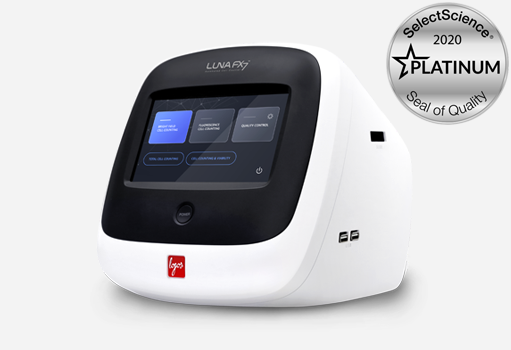
LUNA-FX7 Automated cell counter
LUNA-FX7 Automated cell counter is our most powerful cell counter with autofocusing, dual fluorescence and brightfield detection plus 21 CFR PART 11 compliance.
| Manufacturer | Logos Biosystems |
|---|---|
| Product Series | LUNA |
| Measurement principle | Microscopy - Cell image analysis |
| Application | Live cell image analysis |
| Cell size range | 1 to 90 μm |
| Optics | Brightfield, Dual Fluorescence |
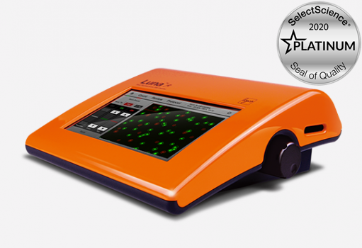
LUNA-FL Dual Fluorescence cell counter
Logos Biosystems LUNA-FL with dual fluorescence and brightfield optics accurately determines cell count, cell viability and GFP transfection efficiency without being limited by cell type or size.
| Manufacturer | Logos Biosystems |
|---|---|
| Product Series | LUNA |
| Measurement principle | Microscopy - Cell image analysis |
| Application | Fluorescence Imaging, Live cell image analysis |
| Cell counting time | 30 sec |
| Cell size range | 1 to 90 μm |
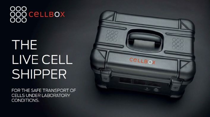
CellBox Live Cell Shipper
The CellBox portable CO2 incubator allows the transport of live cell cultures, tissue and other cell-based samples without the need for freezing, thawing and recovering cells - samples arrive ready to use!
| Manufacturer | Cellbox Solutions |
|---|---|
| Product Series | Cellbox |
| Measurement principle | Microscopy - Cell image analysis |
| Application | Live cell image analysis |
| Incubation temperatures | 28-38°C |
| CO2 Concentration | 0-20% |
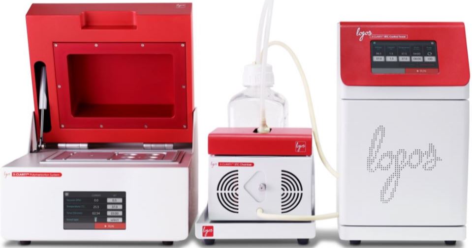
Logos Biosystems X-Clarity
The X-CLARITY is an all-in-one system and ready-to-use reagents for simple, rapid and efficient tissue clearing to allow for 3D analysis of the anatomical structure of tissues.
| Manufacturer | Logos Biosystems |
|---|---|
| Product Series | X-CLARITY |
| Measurement principle | Microscopy - Cell image analysis |
| Application | Fluorescence Imaging, Live cell image analysis |
| Type | Tissue clearing system |
| Throughput | Up to 768 samples |
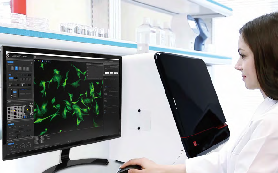
Logos biosystems CELENA X
Automated high content cell imaging and analysis with multicolour fluorescence for drug discovery and cell biology
| Manufacturer | Logos Biosystems |
|---|---|
| Product Series | CELENA |
| Measurement principle | Microscopy - Cell image analysis |
| Application | Live cell image analysis |
| Imaging modes | 4-channel fluorescence, brightfield, phase contrast, colour brightfield |
| Camera | Monochrome and colour CMOS |

Phasefocus Livecyte
Livecyte enables quantitative, label-free live cell imaging with automated tracking and behavioural analysis of hundreds of individual cells within heterogeneous cell populations.
| Manufacturer | Phasefocus |
|---|---|
| Product Series | Phasefocus Livecyte |
| Measurement principle | Microscopy - Cell image analysis, Ptychography |
| Application | Fluorescence Imaging, Live cell image analysis |
| Environmental Control incubator | Full heat, CO2 and humidity control plus sample pod |
| Transmission inverted microscope | 4x to 40x objectives (additional objectives available), 650nm diode laser, sCMOS camera |
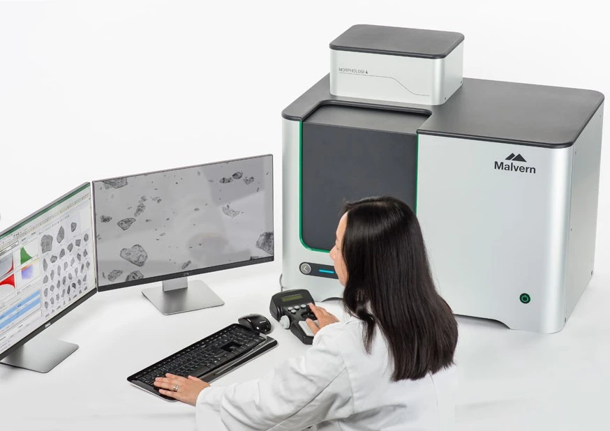
Malvern Morphologi 4
Malvern Morphologi 4 rapidly measures particle size, shape, transparency, count and location of dry powders, fibres and wet suspensions using automated static imaging analysis.
| Manufacturer | Malvern Panalytical |
|---|---|
| Product Series | Malvern Morphologi |
| Measurement principle | Microscopy - Image Analysis |
| Application | Particle Shape, Count & Chemical ID |
| Particle size range | 0.5µm to >1300µm |
| Particle size parameters | Circle equivalent diameter, length, width, perimeter, area, max distance, sphere equivalent vol, fiber total length, fiber width |
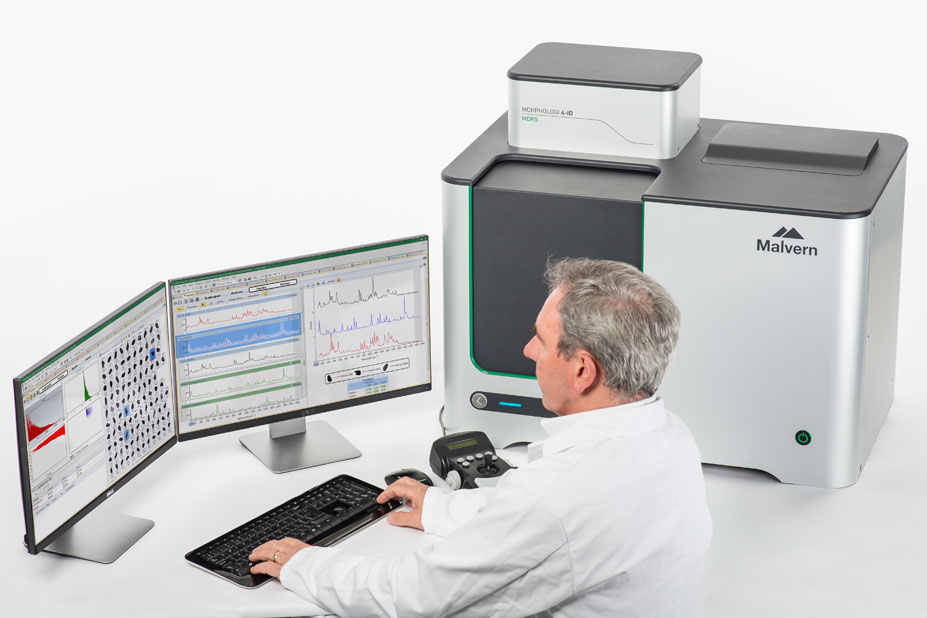
Malvern Morphologi 4-ID
Malvern Morphologi 4-ID combines automated static imaging for particle size, shape, count with Raman Spectroscopy to enable additional chemical identification analysis.
| Manufacturer | Malvern Panalytical |
|---|---|
| Product Series | Malvern Morphologi |
| Measurement principle | Microscopy - Image Analysis + Raman Spectroscopy |
| Application | Particle Shape, Count & Chemical ID |
| Particle size range | 0.5µm to >1300µm |
| Particle size parameters | Circle equivalent diameter, length, width, perimeter, area, max distance, sphere equivalent vol, fiber total length, fiber width |
Customer Installations
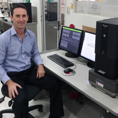
UWS, Advanced Materials Characterisation Facility (AMCF) have installed their new bench-top Phenom XL Scanning Electron Microscope (SEM).
Phenom XL Desktop SEM
The UWS through collaboration with Phenom-World BV, and ATA Scientific has been able to secure a 2 year placement of the bench-top Scanning Electron Microscope (Phenom SEM)
Phenom GSR SEM
UNSW, School of Chemical Engineering has installed their new KSV NIMA Langmuir-Blodgett Trough
Biolin-KSV NIMA LB Trough
LaTrobe University, School of Molecular Sciences, have installed their new NTA system to investigate extracellular vesicles (exosomes, microvesicles)
Malvern NanoSight NS300
SEM & Imaging
A scanning electron microscope (SEM) can be used across a number of industrial, commercial, and research applications. It’s a type of microscope that produces images of a sample by scanning the surface with a focused beam of electrons. The electrons interact with the atoms in the sample, producing various signals that contain information about the surface topography, morphology and composition of the sample. Various types of signals are produced, including secondary electrons (SE), reflected or back-scattered electrons (BSE), characteristic X-rays and light (cathodoluminescence) (CL), absorbed current (specimen current), and transmitted electrons.
SEMs have become a powerful and versatile tool for material characterisation. This is especially so in recent years due to the continuous shrinking of the dimension of materials used in various applications.
SEM & imaging techniques
There are a number of different SEM & imaging techniques, with three of the major ones discussed below:
Static image analysis
In particle characterisation, a distinction is made between static and dynamic image analysis. Dynamic image analysis is ideal for the routine analysis of bulk solids. With high sample throughput and low susceptibility to errors, this method is a great alternative to conventional sieve analysis.
Static image analysis is generally used for narrow size distributions, with a focus on the precise characterisation of mostly fine particles. This method acquires high-resolution particle images that allow for size and shape measurement with the highest level of accuracy. Static image analysis is based on a microscopic procedure in which a slide is photographed step by step and the particle images evaluated automatically.
Ptychographic quantitative phase imaging
Phase imaging techniques are an invaluable tool in microscopy for quickly examining thin, transparent specimens.
Quantitative phase imaging detects minute changes in phase when light propagates through the cell morphology, and it has become the most common approach for fine cell structure distinction without employing higher radiation powers thereby reducing phototoxicity.
Ptychography employs a technique that uses the intensity of light that is scattered, or diffraction patterns of light created, to produce a pattern on a CCD camera. This scattering pattern of light is passed through the Ptychography algorithm to build a high contrast image. It’s recognised as a unique technique, offering several advantages over traditional QPI. Collecting these scattering patterns and processing them through the algorithm elucidates many advantages over traditional microscopy, namely post acquisition focus, high contrast images and minimal light damage to the sample.
Desktop SEM
Desktop SEM provides direct access to the high-resolution and high-quality imaging and analysis required in a large variety of applications. This user friendly tool bridges the gap between the optical microscope and ultra high-resolution electron microscopes. The Phenom Pro Desktop SEM, for example, exceeds the resolution of optical microscopes and eliminates the expense, delay and difficulty associated with operating a traditional SEM. With an integrated X-ray analysis system, sample structures can be physically examined and their elemental composition determined.
Instruments used in SEM & imaging
There are a number of different instruments used in SEM & imaging, including the following:
- Malvern Morphologi 4
- Malvern Morphologi 4-ID
- Phasefocus Livecyte
- Phenom Pharos Desktop SEM
- Phenom GSR Desktop SEM
- Logos Biosystems Celena X
- Logos Biosystems LUNA FX7
Application areas of these instruments include immunological, neurobiological, cancer and basic cell biology research.
ATA Scientific and SEM & imaging
At ATA Scientific our suppliers include some of the world’s most well-regarded companies such as Malvern Panalytical, Phenom World (now part of Thermo Fisher Scientific), Avestin, Phasefocus, Logos Biosystems, Cellbox Solutions and Micromeritics.
Our people have an extensive range of skills and experience in analytical technologies, especially particle and protein characterisation. As well as supplying modern SEM and imaging instruments, we can support you with ongoing applications assistance and encouraging the adoption of standard operating procedures. Advice regarding instrument operation and optimisation of data quality are freely available by phone and email.
If your research or scientific organisation is looking for the world’s best laboratory equipment and expert support for SEM & imaging, contact us to discuss your requirements.
Looking for the most suitable analytical system for your project?
Call us directly on 02 9541 3500 for a free consultation, browse our extensive range online or find the right instrument with our easy to use Product Finder
Browse product range Request FREE Consultation