


 02 9541 3500
How to make Cryo EM more available to researchers
Back in 2017 Jacques Dubochet, Joachim Frank and Richard Henderson had won the 2017 Nobel Prize in chemistry for their work developing cryo-electron microscopy technology – or cryo-EM. The technique allows images to be taken of protein components that are reconstructed into movie like scenes that show how biological machines work. It generates atomic resolution 3D models of molecules that are not able to be seen using other structural biology techniques to gain insight into things like receptors that are therapeutic drug targets, molecular motors that deliver cargo to different parts of the cell and emerging viruses that lead to human disease.
While X-ray crystallography is widely known and used as the gold standard for generating high-resolution biomolecular images, this older technique requires the formation of ordered crystals. Proteins must first arrange together in repeating patterns which can be challenging. With cryo-EM, there’s no need for biomolecules be ordered in this way.
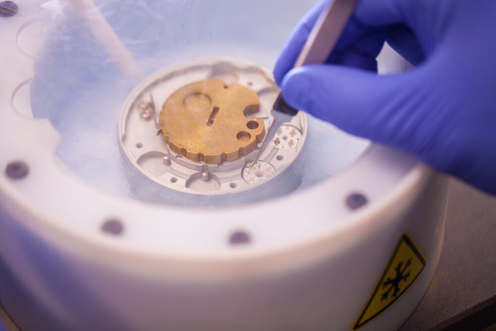
Cryo EM saves time, but it can be very expensive and requires massive amounts of computational power and data storage, so many of these technologies are only available at major microscopy facilities.
In addition, sample preparation for cryo-EM can be a demanding and expensive process, requiring several rounds of purification and screening to isolate the macromolecules of interest. Negative stain electron microscopy (EM) is one of the established tools for the initial screening of purified samples prior to imaging using transmission electron microscope (TEM) to study particle size, morphology, concentration, and agglomeration. With the introduction of the desktop scanning transmission electron microscope (STEM), an alternate process for sample screening has been developed that is faster and more cost-effective than the traditional methods.
So, the question is, how does a desktop STEM system help enable the specialised and expensive resources required for cryo-EM to be more broadly available to researchers across the country and around the world?
What is STEM?
Scanning transmission electron microscopy (STEM) is a high-resolution imaging technique used to visualise structure and composition of materials and biomolecules. It scans a focused electron beam across a thin sample collecting signals based on scattering angle to produce bright field (phase contrast) or dark field (compositional contrast) images. When paired with energy-dispersive X-ray spectroscopy (EDS) it can provide elemental composition.
Samples for STEM imaging need to be electron transparent and typically 100 nm thickness or less to minimise scattering and improve signal to noise.
What is the difference between STEM and TEM?
In an SEM, the secondary electron (SE) and backscattered electrons (BSE) are used to acquire images of a sample’s surface whereas in a TEM, the transmitted electrons are detected to produce a projection-image through a sample’s interior. STEM can be thought of as a hybrid between SEM and TEM, where a thin sample is scanned with a focused electron beam (like an SEM) and the transmitted electrons are detected (like in TEM) at each point to acquire a high-resolution image of the internal structure of the sample.
Why use STEM-IN-SEM?
When compared to traditional TEM techniques, integrating STEM-in-SEM provides several advantages which is driving its growth in popularity. The resolution meets most application’s needs and produces results much faster than it can be obtained using a TEM, improving workflow throughput significantly. Additionally, SEMs operate at low acceleration voltages which can improve imaging contrast (especially for biological samples like carbon nanotubes or biological tissue) and causes less beam damage to the sample enabling longer working windows for delicate samples. STEM structural information can be easily correlated with BSD and SED images typically provided using SEM. The technique is also much less sensitive to sample thickness and is easier to prepare than TEM, meaning thicker, uneven and unstained samples can be quickly and easily imaged.
Sample preparation techniques used in Electron Microscopy
Two techniques are commonly used – negative and positive staining.
First developed in the mid-twentieth century, the negative staining technique uses a protocol for staining virus particles with heavy-metal salts and observing them in a TEM
Similarly, the positive staining technique has been used since the late 1950s for enhancing contrast of biological samples (tissues and cell structures, viruses etc). Thin sections of samples fixed in glutaraldehyde and embedded in epoxy resin are placed onto copper grids and incubated in heavy metal salt solutions that react with cell structures before imaged using a TEM. Positive staining results in a small amount of stain forming a thin shell around the molecule, meaning the sample appears dark against a light background.
A conventional TEM is generally used to visualise the stained samples. This results in expensive equipment being tied up in screening samples rather than their intended application in high-resolution imaging. The facility costs and frequent maintenance costs can be significant, imposing financial burdens on principal investigators who are relying on it for sample screening. The learning curve on how to use a TEM can be rather steep with new users taking several days to weeks to become proficient enough to operate a TEM independently. Therefore, exploring alternatives to dedicated TEM instruments is an appealing choice for those who want to maximse the benefits of stain EM for sample screening.
The Desktop Phenom Pharos FEG- SEM with STEM detector
Desktop Phenom Pharos FEG-SEM is a compact, easy to use imaging system that can be used as a quick imaging tool that complements and helps free up valuable time on larger, more complex and much more expensive EM imaging systems.
When equipped with the STEM detector Phenom delivers ultrastructural characterisation with resolution close to a TEM, but with a larger field of view. The resolution meets most application’s needs and its ease of use allows images to be generated by user of any experience level in as little as one minute from sample loading. This new imaging modality provides immense value to a variety of fields including nanomaterials, catalysts, pathology, and batteries.
The Phenom Pharos Desktop SEM with a STEM detector is a plug-and-play accessory that integrates a segmented solid-state detector into a sample holder. It is compatible with standard EM grids and is capable of independent collection of bright-field (BF), annular dark-field (ADF), and high-angle annular dark-field (HAADF) signals.
Options for STEM imaging
Phenom SEM can be programmed to collect data in an automated fashion, allowing for quick inspection of the entire grid. This allows users to very quickly and easily collect images containing hundreds of thousands of particles that can be used for statistical analysis.
The sample holder is compatible with standard 3 mm TEM grids. A clamp-based mount ensures that delicate samples are safely loaded and securely held in place during handling. The STEM detector and its electronics are conveniently integrated into the holder. Switching between conventional SEM and STEM imaging modes is as simple as loading a new sample. Users can obtain a time-to-image of less than 40 seconds and select from three standard STEM imaging modes; bright field (BF), dark field (DF) and high angle annular dark field (HAADF); or explore custom configurations.
Bright Field (BF)
BF imaging collects on-axis electrons scattered by the sample. Contrast depends primarily on sample thickness and composition, where thicker areas containing heavier elements appear darker. With improved sensitivity to light elements, BF mode can be particularly useful for studying organic samples. For example, when investigating carbon nanotubes, the SED image may not be able to easily locate metallic catalyst particles, however by using the BF STEM on the Pharos SEM these particles are clearly shown as dark spots.
Dark Field (DF)
DF imaging detects off-axis electrons that result from relatively lower diffraction angles. Image contrast depends on thickness and atomic number with brighter areas corresponding to local mass-thickness. DF imaging is more sensitive to atomic number differences in lighter elements and is useful for a broad range of samples.
High Angle Annular Dark Field (HAADF)
HAADF imaging collects the off-axis signals at the highest scattering angles and is most sensitive to atomic number contrast, or Z contrast. HAADF is particularly sensitive to heavier elements such as metal atoms. This mode can be used to detect features that are harder to visualise with the other imaging modes.
Collaboration with NSW Health Pathology and the Ingham institute for Applied Medical Research
Electron microscopy plays a major role in diagnosing renal and rare diseases, however high costs can limit its access. Professor Murray Killingsworth from NSW Health Pathology’s Liverpool lab and Dr Tzipi Cohen Hyams from the Correlative Microscopy Facility at the Ingham Institute for Applied Medical Research are working with ATA Scientific and Thermo Fisher Scientific International to develop the Phenom Pharos FEG-SEM with low kV STEM imaging and assess how it may be used for high resolution ultrastructural characterisation of soft tissue and cells for cell biology and pathology. The team recently won “Recognising our Pioneering Spirit” Award for their pioneering work at the 2023 NSW Health Pathology Awards! The group will also be presenting their latest findings and demonstrating the Phenom Pharos together with ATA Scientific during the UltraPath XXI conference 2024 to be held in Sydney (September 30th – October 4, 2024).
If you are eager to explore the capabilities of the Phenom Pharos FEG-SEM, we are delighted to offer you a personal demonstration. Request a guided demo using your own samples with a product specialist.