


 02 9541 3500
An Introduction to the Microscopy Facility Toolkit
Building a Microscopy facility must be a daunting task, with such an array of imaging technologies and applications. Gone are the days of the humble compound microscope whose predecessor illuminated the ‘cell’ as coined by Robert Hooke1. I can only imagine the excitement of such a discovery and ponder how many times microscopy has yielded such emotion since then. Microscopy unlocks a window into a wondrous world, there is no debate around its value, just what is the most useful.
It is overly simplistic to consider that there need only a few technologies to fulfil the gambit of application needs. As with most things, it’s a function of ‘horses for courses’ as one technology is great at specific functions though not so fantastic at others. As technology marches on, the shiny new ‘must have’ attracts the researcher desperate to publish novel findings elucidated by a magical black box developed by a clever physicist. This hunger is fed by the perennial ‘publish or perish’ mantra exacerbated by the anecdotal tendency for novel technology to gain favourable acceptance by journals. The issue of competing interests should not be overlooked.
The Arms Race
The arms race in microscopy is not just driven by the researcher. Consider the microscope manufacturers desire, a FOMO, adding their flair to a particular development they have not invented but want to have a me-too with added ‘bells and whistles’ to gain the upper hand in a sale and capitalising on brand loyalties. Super-resolution microscopy is a great example of this, by delivering optical images with spatial resolutions below the diffraction limit, several super-resolution fluorescence microscopy techniques have opened new opportunities to study biological structures with details approaching molecular structure sizes2 – note STED, SSIM , PALM, STORM and RESOLFT. Bending the frontier delving beyond what was thought impossible. Beautiful discoveries, however, it still has limitations in capabilities.
A divergence is occurring, a whole new world of computational microscopy is emerging to enhance the image we no longer see. This concept is spawning a plethora of variations on the theme in the race to the ultimate in resolution. Whilst many are all-consumed by this resolution race, others are inspired by not just the individual, but the population – the company they keep in equal measure. We can all plead guilty to only seeing the majority and overlook the outlier – visualise a cluster in a flow cytometry experiment and consider the gating applied to the data along with the premise of these actions. This may well be the cell that is going to kill the patient.
Why bother?
Perhaps it is time we consider a better way of investigating the behaviour of live cells – a novel method of looking at the cells in question that has a very large field of view to enhance the statistics, a way to view them for extended time frames, even weeks, without perturbing, killing, or bleaching. A gentle nourishing environment to keep cells happy and label-free is ideal. You may be thinking – Why bother given there are so many options for microscopes? In short, signal to noise ratio is limited when using standard microscopy methods. While the ubiquitous use of fluorescence may seem to have solved this, new data is emerging that, arguably and at times unknowingly, has exposed fluorescence as a major influencer of cell behaviours itself. Rarely can you gain such powerful insight with incredible single cell tracking and metrics right through to entire populations using Fluorescence alone.
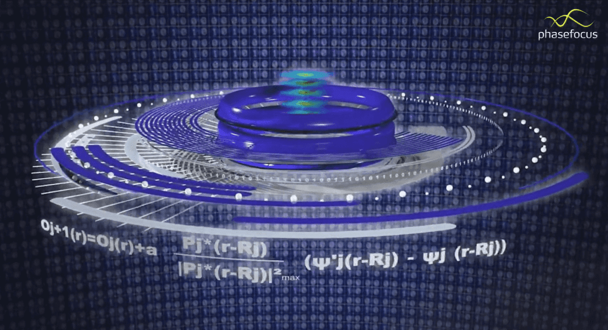
How does Ptychography work?
Ptychography is not only an unusual name, but an unusual technology. It is a hybrid of sorts, a blend between an optical microscope and a light scattering detector. Briefly, the optical microscope sends light through a sample and a scatter or diffraction pattern is received on the detector, a digital method is used to translate the diffraction pattern into a quantitative image this is the Phasefocus virtual lens. The patterns are processed by the Ptychography algorithm to produce quantitative intensity and phase images of the sample. To explore this further please click here3.
The resultant is a dramatic increase in the Signal: Noise giving the opportunity to avoid fluorescent probes, enabling a very low intensity light source avoiding cellular damage. This is a big win for the cells and the researcher trying to see them in an environment as natural as possible.
A Bitter Pill
It is tough to acknowledge that the core method of Fluorescence employed by so many in cell biology may be delivering flawed results. Several studies run on cells in parallel with and without a fluorescent label have shown profound changes to behaviours and other indicators such as proliferation, motility, and dry mass, prompting the question – Why aren’t more facilities using this technology if only for a confirmatory application notwithstanding the enormity of insight it offers beyond this? One could postulate it may be because users are blinded by resolution.
What am I likely to see in a facility?
Summarising the breadth of microscopes with their strengths and weaknesses is not a simple task and in doing so assumptions must be made along with the lens we look through. The image below gives a summary, but more importantly, it shows where Livecyte fits into the scheme of a broader microscopy facility. It is worth noting, Livecyte does not dispense with any, it enhances the offering within a facility.

To gain a little insight into cells doing weird things I encourage you to link up with the Phasefocus Twitter feed https://twitter.com/PhaseFocus1
Further to this, please contact us at ATA Scientific. We will be happy to introduce you to some systems, perhaps arrange a demonstration. Call us on +61 2 9541 3500 or send an email to pdavis@atascientific.com.au
References
- https://www.nationalgeographic.org/encyclopedia/cell-theory/ site accessed 1Feb2022
- Godin AG, Lounis B, Cognet L. Super-resolution microscopy approaches for live cell imaging. Biophys J. 2014;107(8):1777-1784. doi:10.1016/j.bpj.2014.08.028
- The Virtual Lens, Phasefocus. https://www.phasefocus.com/technology Website accessed 7 Feb2022.