


 02 9541 3500
Live cell imaging and analysis using Quantitative Phase Imaging (QPI)
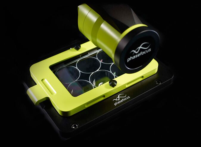
ATA Scientific is pleased to be the new local distributor for the Livecyte™ Cell Imaging and Analysis system developed by UK-based company Phasefocus™.
The Livecyte is a unique imaging system for live cell analysis used to study cell functions and behaviour. It can reveal the inner details of transparent structures without the need for staining or tagging. Based on Ptychographic Quantitative Phase Imaging (QPI) technology, individual cells and cell populations can be automatically tracked and analysed for phenotypic and kinetic behaviour including motility over several hours or days.
A large field of view ensures that highly motile cells are not “lost” during long time-courses. High contrast images and videos can be generated which are artefact free and quantitative in the absence of labels or high intensity light imaging which can potentially disturb normal cell functions.
With the ability to automatically analyse multiple parameters simultaneously from a single experiment, scientists can accurately and efficiently define the impact of experimental conditions on each and every cell. This makes the Livecyte pertinent across a wide range of application areas including immunological, neurobiological, cancer and basic cell biology research.
For further details click here.