


 02 9541 3500
Under the Microscope: Key Differences Between SEM and Optical Microscopy
In the field of scientific imaging, operators typically rely on optical and scanning electron microscopes. While both instruments help characterise microscopic samples, they do so in very different ways.
Optical Microscopy
Optical microscopes are the ‘traditional’ type of microscope in that they use a lens to produce a magnified image. For scientific uses, optical microscopes can be quite complex, however the underlying principle remains the same.
Optical technology
Optical microscopes possess two major functions:
- production of a magnified image of a sample of specimen
- illumination of a specimen
Optical microscopes share some similar components as other optical instruments like binoculars and telescopes. For example, each of these optical instruments has either:
- converging lens
- concave mirror
In optical microscopes, you will more commonly find a converging lens. The presence of a concave mirror is used mostly for illumination of the sample, similar to how headlights work in cars.
So, we see how the primary purposes of an optical microscope, to magnify and illuminate, are in part achieved through the use of common optical components in the converging lens and concave mirror.
Forming an image through the lens
Even if you’ve never engaged with an optical microscope scientifically, you have probably used one casually. For example, many of us have used a handheld magnifier to create a heat point on the ground, probably as children.
This shared experience helps us to understand two significant terms in optical lens magnification.
- focal point
- focal length
Focal point
As in the example above, the point of heat when magnifying light onto the ground is the focal point.
Focal length
The focal length is the distance from the lens to the focal point.
Focal length and curvature of lens
In optical magnification, there is a relationship between the focal length and the curvature of the converging lens. The smaller the radius of curvature the shorter the focal length. Related to this, a lens with a large diameter is found to be more effective.
Numerical aperture
In the study of microscopy, the relationship between diameter of the objective lens and focal length is called Numerical Aperture or NA for short.
According to Leica Microsystems:
“NA = n sin α, where n is the refractive index of the medium filling the space between the object and the lens, and α is the half-angle of the maximum cone of light that can enter the lens.”
Scanning Electron Microscopy
Scanning electron microscopy uses a beam of focused, high-energy electrons to generate surface signals on solid samples.
These signals reveal information about the:
- texture (sometimes called morphology)
- chemical composition
- crystalline structure
- material orientation
This can be rendered in a two dimensional image, with most SEMs able to scan and image materials from 1cm to 5 microns in width.
Kinetic energy and dissipation
Electrons accelerated by the SEM produce significant kinetic energy. When they ‘interact’ with a sample they dissipate a number of different signals. This is known as Electron-sample interactions. The production of these signals includes:
- secondary electrons which produce the SEM image
- backscattered electrons which also produce the image
- diffracted backscattered electrons which determine orientations of minerals and crystal structures
- photons used for elemental analysis and continuum x-rays
- visible light
- heat
Creating a SEM image
As demonstrated, secondary electrons and backscattered electrons produce SEM images as a result of electron-sample interactions.
Secondary electrons are more valuable for showing morphology and topography, while backscattered electrons are useful for illustrating composition contrasts in multiphase samples.
Key differences between Scanning Electron Microscopy and Optical Microscopy
Ease of use
Optical Microscopy is attractive in no small part thanks to the ease of operation. With Optical Microscopy a sample can be:
- analyzed in air or water
- imaged in natural colouring
This makes Optical Microscopy an attractive solution when compared to traditional large SEM’s typically found in University microscopy centers.
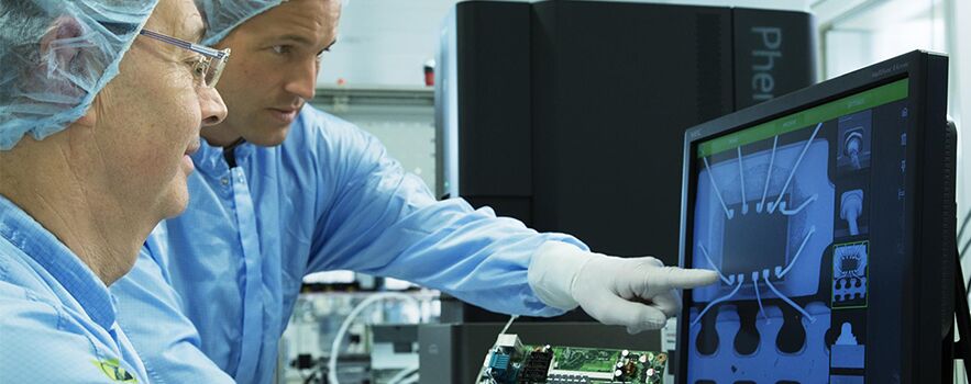
However, recent developments in SEM imaging in particular the new desktop Phenom SEM series of instruments offer users an affordable and easy to use imaging tool that bridges the gap between the optical microscope and ultra-high resolution microscopes. The unique optical navigation camera displays a view of the entire sample and allows the user to move to any spot on the sample with just a single click. The proprietary venting/loading mechanism supports the highest throughput even for large samples up to 100mm x 100mm, and ensures a time-to-image of less than 60 seconds.
Resolving power
In microscopy, the resolving power is essentially the smallest ‘detail’ that can be resolved or seen by the microscope. Resolving power is directly influenced by the wavelength of the imaging beam used by the microscope.
Wavelengths of SEM and Optical Microscopy light
Because the resolving power of an Optical Microscopy is limited to visible light, the wavelength of the beam is 400-700 nanometers. Therefore optical microscopes can only offer upto 1,500 x magnification and may not go below 200 nm in resolution.
In contrast, the light beam used by a SEM is comprised of energies up to a thousand times greater than visible light, the depth of focus and resolving power in an SEM instrument is much greater, providing a significant advantage.
The Phenom SEM has a magnification range up to 150,000x and resolution of <8nm and thus delivers more detailed information. A long-life CeB6 electron source in combination with the four-segment Backscatter detector (BSD) yields sharp images together with chemical contrast information. The Phenom can also be fitted with a Secondary Electron Detector (SED) that supports surface sensitive imaging. Unlike other systems they include a fully integrated X-Ray analysis (Energy Dispersive Spectrometer, EDS) that allows the user to quickly identify and assess the distribution of elements in a sample.
Which microscopy instrument is best for you?
Both microscope types have their advantages and drawbacks. SEM’s greater depth of focus and a high resolving power draw you one way, while ease of use and maintenance of the optical microscope draw you the other.
The Desktop Phenom SEM series of instruments offer users a fast and easy to use electron microscope with high resolution imaging and element identification – all advanced features that may not be available when using an optical system.
Depending on your use case, you may already know which type of instrument is right for you. Browse our full range of SEM and Optical Microscopy instruments today, or contact ATA Scientific for a quote.